


Dr. Shumin Duan/'s group published in the Jounal of Neuroscience
A study from Professor Shumin Duan’s group on how an intracellular size filter enhances antigen processing in microglia was recently published on The Journal of Neuroscience
Abstract:
Microglia are the primary immune cells in the brain. Theycontinually survey the brain microenvironment and uptake cell debris and aberrant proteins to maintain brain homeostasis. Recent studies have shown that reactive microglia are able to present antigens to T cells and initiate immune responses. However, how the uptaken antigens were processed, trafficked and presented to T cells remains a mystery.
In a study recently published on The Journal of Neuroscience, Cong and Huiquan, two PhD students in Duan’s group, elegantly addressed this question by using pulse-chase labeling, coupled with real-time fluorescence confocal microscopy and magnetic nano-bead assays. They have found that activated microglia engulf a large amount of soluble antigens by pinocytosis. Then, the engulfed contents were hydrolyzed and selectively transferred to different cellular compartments via a size-based sorting (size filter) mechanism: the smaller hydrolyzed peptides were preferentially delivered to the major histocompatibility complex class II (MHC-II) containing lysosomes, whereas the non-hydrolyzed proteins remain within the pinosomes. It appears that transient fusion events between pinosomes and lysosomes mediate this selective transfer in a Rab7 and dynamin II-dependent manner. Disrupting the size-based sorting in microglia markedly reduced the proliferation and cytokine release of co-cultured CD4+ T cells, suggesting that size-based sorting enhances antigen processing and presentation. These findings reveal a novel sorting mechanism for soluble internalized contents in microglia and provide important new insights intothe evoked immune responses observed with a wide range of brain disorders in which microglia activation are involved.
This study has raised extensive interests and was selected by the editors as a featured article in the issue (http://www.jneurosci.org/content/35/6/i.short). This study won’t be possible without supports from the Major State Basic Research Program of China and , the National Natural Science Foundation of China., and collaboration with multiple labs (Drs. Li YU, Linrong LU and Xuetao CAO).
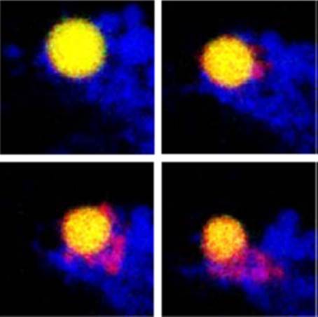
Figure 1. Time-lapse images of a microglial cell induced to undergo pinocytosis in the presence of large (green) and small (red) dextrans. Initially, the dextrans were enclosed in the same pinosome ( yellow, top left),which was surrounded by lysosomes (blue). After 8 min, smaller dextrans had begun to move into lysosomes (top right). More small dextranss had exited the pinosome 2 and 4 min later (bottom left and right), leaving the larger dextrans behind.
